


Dental X-Rays Radiation From Dental X Rays Are Safer Than They Used To Be |
Posted: August 17, 2017 |
A standout amongst the most imperative instruments family dental specialists utilize is the dental x-beam. X-beams enable dental specialists to decide conditions that may not be apparent amid visual examination. Shrouded conditions, for example, root sickness, affected teeth and issues of the jaw can be found. Issues that may influence fruitful revival of teeth can be recognized by restorative dental specialists. Today, both x-beam gear and methods have been refined to an abnormal state of exactness that makes them more secure than at any other time. History of X-Rays in Dentistry The historical backdrop of dental x-beams starts with the disclosure of radium by Wilhelm Conrad Roentgen in 1895. The capacity to see the boney structures inside the body opened up new chances to comprehend and treat various infections and wounds. Soon after this disclosure, Dr. Otto Walkhoff in Braunschweig, Germany, took the primary x-beam of the oral cavity. These early x-beams set aside fundamentally greater opportunity to create a picture, regularly as long as 25 minutes. The new innovation rapidly spread all through Europe and to the United States, where it was utilized to help patients with shrouded dental conditions. Proposals on Dental X-beams On account of the threats of unreasonable measures of radiation amid indicative x-beams, strict controls have been put on the utilization of x-beam hardware. Organizations, for example, the U.S. Nourishment and Drug Administration, and additionally the American Dental Association, gives various rules with respect to who ought to get dental x-beams, what number of x-beams ought to be controlled and the radiation from dental x rays most secure approach to regulate x-beams. These proposals are actualized to guarantee that base measures of radiation are utilized to analyze dental issues.
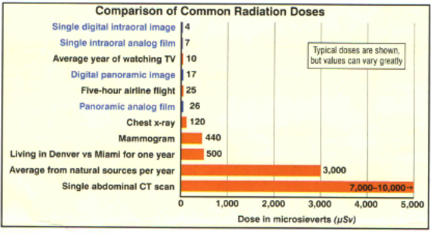
Safe Levels of Radiation Radiation happens normally in the earth, and individuals are presented to it as they approach their ordinary lives. A few exercises, for example, taking a plane flight, open individuals to somewhat more elevated amounts of radiation. The measure of radiation patients get because of dental x-beams is a great many circumstances not as much as common radiation. Sadly, there is no known "safe" level of radiation. As a result of this vulnerability, restorative and dental hardware originators constantly endeavor to create gear that utilizations less radiation than past gear, yet still successfully makes the pictures that assistance to analyze and treat medicinal and dental conditions. Dental X-Rays Have Improved In the beginning of x-beams, patients were presented to a lot of radiation that could conceivably cause growth numerous years after the fact. Today, new innovation has permitted x-beam hardware to utilize minute measures of radiation to deliver fantastic pictures of the structures within the teeth and jaw, with negligible hazard. Computerized radiography utilizes around 90 percent less radiation than conventional film x-beams. The computerized hardware likewise takes out the requirement for chemicals to process the pictures, making them all the more ecologically agreeable. PC tomography, or CT filters, that permit the considerably more prominent examination and conclusion of inward structures conceivable, utilizing little measures of radiation. Advances in Dental X-Ray Technology The hand-held x-beam machine and different advancements are conveying the energy of radiologic conclusion to territories of the world that already couldn't bolster this unpredictable innovation. Presumably more noteworthy upgrades will convey significantly more security and accommodation to x-beam gear later on.
|
||||||||||||||||||
|
||||||||||||||||||